Po-Cheng Kung, Jian-Min Zuo, Brian Somerday, Petros Sofronis, Jessica Anne Krogstad
Short-range ordering (SRO) in crystalline materials can be categorized in two main groups: homogeneous and heterogeneous SRO [1,2]. Homogeneous SRO refers to a statistical deviation from random that is present continuously and uniformly throughout the sample. On the other hand, heterogeneous SRO consists of nanometer scale domains in which different alloy elements are prone to occupy different lattice sites in an ordered fashion, as opposed to a randomly distributed solid solution. The common size of heterogeneous SRO domains is 2 to 5 nm [1], thus microscopy techniques with spatial resolution of nanometers are required to characterize individual heterogeneous SRO domain.
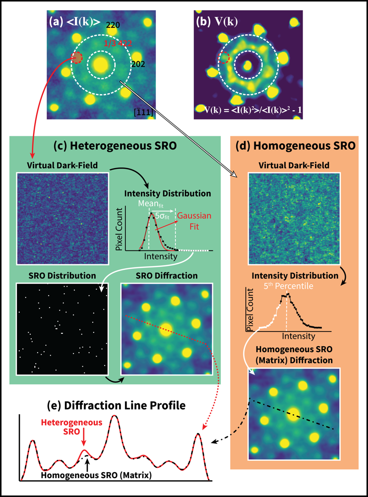
Among popular microscopy techniques, we believe the Scanning Electron Nano-Beam Diffraction (SEND), or 4D-STEM, is most suitable for characterizing SRO in alloys. By scanning the electron nano-beam across the specimen and collecting the local electron diffraction pattern at different position, one can map the distribution of SRO domains and study the diffraction behavior of several SROs and the matrix at the same time. Here, we introduce a procedure utilizing the Fluctuation Electron Microscopy (FEM) method that is commonly used in studying amorphous materials [3] to analyze homogeneous SRO signals and identify heterogeneous SRO domains that are statistically different from the matrix. Since the heterogeneous SRO domains are embedded in a homogeneous matrix, we can calculate the normalized variance of weak diffraction intensity in the series of diffraction patterns to differentiate signals caused by small domains (heterogeneous SRO, surface oxide, etc.) from the ones stem from the matrix.
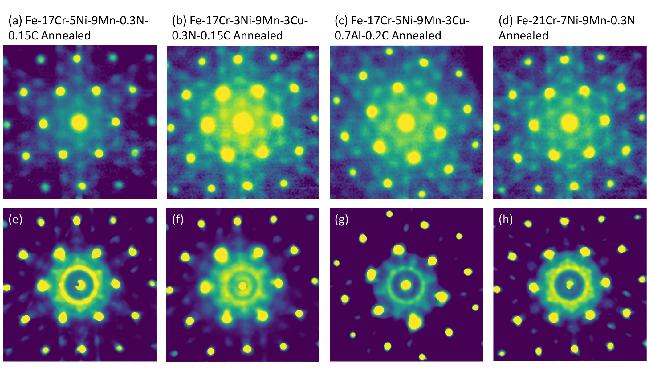
Figure 1 presents an example of the proposed procedure on a solution treated Fe-17Cr-5Ni-9Mn-0.3N-0.15C austenitic steel. We show that the 1/3{422} weak reflections in [111] zone seen in the averaged diffraction pattern, which are commonly assigned as SRO diffraction signals in FCC alloys [4,5], are caused by three different features in the material: homogeneous SRO in the matrix, isolated heterogeneous SRO domains, and surface oxides. We are not only able to show the existence and distribution of different types of SRO, but also detect minute differences in the SRO weak diffraction, which are essential for ultimately solving the ordered structure. Figure 2 is composed of the diffraction patterns and variance maps of annealed Fe-17Cr-5Ni-9Mn-0.3N-0.15C and a few other similar alloys. While the averaged diffraction patterns are nearly identical for all alloys, the variance maps reveal the changes in heterogeneous SRO diffractions from sample to sample. These differences hint at how SRO structure and morphology changes when altering the alloy composition and heat treatment history. In this poster presentation, we will demonstrate how this procedure applies to austenitic steels in detail, while also addressing further potential applications and acknowledging limitations of the method.
Reference:
1. Schönfeld, B. Local atomic arrangements in binary alloys. Progress in Materials Science 1999, 44, 435–543.
2. Owen, L. R.; Playford, H. Y.; Stone, H. J.; Tucker, M. G. A new approach to the analysis of short-range order in alloys using total scattering. Acta Materialia 2016, 115, 155–166.
3. Voyles, P. M., and D. A. Muller. “Fluctuation microscopy in the STEM.” Ultramicroscopy 93.2 (2002): 147-159.
4. Kim, Y. S.; Maeng, W. Y.; Kim, S. S. Effect of short-range ordering on stress corrosion cracking susceptibility of Alloy 600 studied by electron and neutron diffraction. Acta Materialia 2015, 83, 507–515
5. Kim, Y. S.; Kim, S. S.; Choe, B. H. The role of hydrogen in hydrogen embrittlement of metals: the case of stainless steel. Metals 2019, 9, 406
Acknowledgments:
This research is supported by Department of Energy Office of Energy Efficiency and Renewable Energy under DE-EE0008832 and was carried out at Materials Research Laboratory Central Research Facilities at University of Illinois at Urbana-Champaign. The alloys studied in this research is provided by Kyushu University and Sandia National Laboratory.